By Dr Beth Watson Bsc DVM
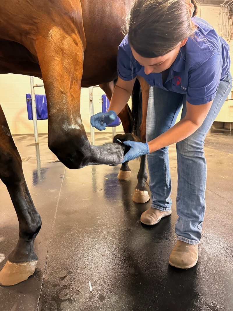
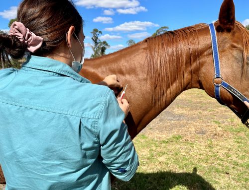
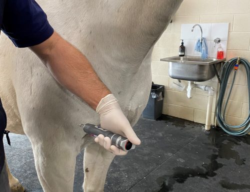
A nerve block is the application of local anaesthetic to a specific region of the horse’s body. Local anaesthetic stops or reduces nerve impulses to the brain. When used as part of a lameness exam vets are aiming to remove sensation from a particular area. If the application of a nerve block changes the lameness or resolves a lameness, we infer that pain in the region of the block is a part of the horse’s presentation. In this way, nerve blocks are used systematically to rule in or rule out areas of the horse’s anatomy to localise and define the lameness.
Nerve blocks for lameness exams are most frequently perineural (around a nerve) or intra articular (into a joint). Perineural nerve blocks involve local anaesthetic being instilled in the facia around the nerve. After a period of time the horse’s limb can be tested for sensation. If it has been effective, the area distal to the nerve block is anesthetised. Care must be taken with interpretation of blocks as there is potential for the local anaesthetic to migrate, altering the pattern of the block and therefore the results. In the case of an intra articular block, the local anaesthetic is instilled directly into the joint. If applied correctly the block should only affect the joint and not the surrounding structures, however if there is deep bone pain or joint instability an intra articular block can provide a false negative. Skilled veterinarians interpret all nerve blocks with care.
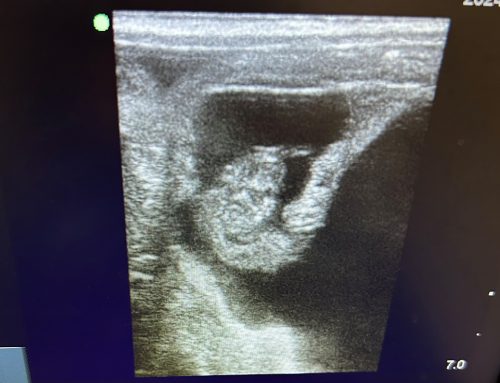
Whilst the lame leg or legs may be relatively easy to identify, it can often be a large jump to diagnose the precise source of the problem, particularly in the case of a chronic lameness. Skilled application of nerve blocks can narrow the problem down to a region. Once a region is identified, this can be further assessed, usually involving diagnostic imaging.
Some might ask why nerve blocks are important if we can use imaging to diagnose pathology in the horse. While this may be true, there is potential diagnostic imaging alone to lead to erroneous conclusions. Since our patients cannot tell us where the pain is, we are making assumptions based on how the horse presents, moves and responds. Human literature suggests in many cases radiological findings are poorly correlated with clinical presentation. While this is more relevant for some disease processes than others, we can extrapolate this to the animal model. This point highlights the need for pain localisation in lameness exams, which is best achieved through the application of nerve blocks. While nerve blocks do not hold all of the answers, they do play a very important part in the complete lameness exam.

